Anatomy Digestive System starting from mouth to excretion.ppt
Anatomy of Digestive system is very helpful well designed ppt to understand how our body works. It also helps to know how disease attacks our digestive system. It contains. ## Description for Anatomy of Human Digestive System PPT **A comprehensive PowerPoint presentation on the anatomy of the human digestive system will provide a visual and informative guide to the structure and function of this vital organ system.** The presentation should include: ### Introduction * Brief overview of the digestive system's role * Importance of understanding its anatomy ### Major Organs and Structures * **Oral cavity:** Teeth, tongue, salivary glands * **Pharynx and esophagus:** Structure and function in swallowing * **Stomach:** Anatomy, including cardia, fundus, body, antrum, pylorus * **Small intestine:** Duodenum, jejunum, ileum, villi, microvilli * **Large intestine:** Cecum, colon (ascending, transverse, descending, sigmoid), rectum, anus * **Accessory organs:** Liver, gallbladder, pancreas ### Detailed Anatomy and Histology * Microscopic structure of different digestive organs * Layers of the digestive tract (mucosa, submucosa, muscularis externa, serosa) * Specialized tissues and cells (e.g., gastric glands, intestinal crypts) ### Visual Aids * Clear diagrams and images of digestive organs * Histological slides or micrographs * Animations or videos to demonstrate processes like peristalsis ### Key Points to Emphasize * Relationship between structure and function * Adaptations of different organs for specific digestive tasks * Blood supply and nerve innervation of the digestive system ### Conclusion * Recapitulation of the major components * Importance of a healthy digestive system **By combining clear explanations, detailed visuals, and relevant clinical information, this PowerPoint presentation will serve as an excellent educational resource for students, healthcare professionals, or anyone interested in human anatomy and physiology.** **Would you like to focus on a specific part of the digestive system, or do you need help with creating the PowerPoint slides?** ## Description for Anatomy of Human Digestive System PPT **A comprehensive PowerPoint presentation on the anatomy of the human digestive system will provide a visual and informative guide to the structure and function of this vital organ system.** The presentation should include: Introduction * Brief overview of the digestive system's role * Importance of understanding its anatomy Major Organs and Structures Oral cavity: Teeth, tongue, salivary glands Pharynx and esophagus: Structure and function in swallowing Stomach: Anatomy, including cardia, fundus, body, antrum, pylorus Small intestine: Duodenum, jejunum, ileum, villi, microvilli Large intestine: Cecum, colon (ascending, transverse, descending, sigmoid), rectum, anus Accessory organs: Liver, gallbladder, pancreas. Read less

More Related Content
- 2. What is the digestive system? The digestive system is a continuous tube that begins at the mouth and ends at the anus. Measuring about 30 feet long in the average adult, it is known as the alimentary canal or gastrointestinal tract. It has 3 functions: the digestion of food into nutrients, the absorption of nutrients into the bloodstream, and the elimination of solid wastes.
- 3. The mouth: salivary glands… Three pairs of glands open into the oral cavity, producing saliva: the parotid (pah RŎD ed), sublingual (sub LIN GWUL), and submandibular (sub man DIB you ler) glands. The sensory organs such as the nose and eyes send a message to the brain, the brain sends the message to the salivary glands, and they secrete the chemicals to begin the digestive process.
- 4. The mouth: tongue The tongue is a muscle covered with a mucous membrane. It has a rear portion called the root, the tip, and the central body. It is covered with taste buds and raised elevations called papillae (pah PILL ah). The taste buds taste sweet, sour, salt, bitter, and umami (oo MAH mee) or savory.
- 5. The mouth: teeth The teeth are used for chewing the food…mastication. The food is broken apart and mixed with saliva to form a bolus, ready to be swallowed. Muscular constrictions move the bolus through the pharynx (soft palate at the back of the mouth) and into the esophagus while blocking the opening to the larynx and preventing the food from entering the airway.
- 6. The esophagus… The food is moved down the esophagus toward the stomach by wavelike muscular contractions called peristalsis. At the opening of the stomach is the lower esophageal sphincter. This is a muscle valve that permits the passage of food, but not the backup of stomach contents under normal conditions.
- 7. The stomach… The stomach is a muscular, expandable organ, the upper portion called the fundus and the lower portion called the antrum. Hydrochloric acid and other gastric juices convert the food to a semiliquid state called chyme. Chyme passes through the pyloric sphincter valve at the bottom of the stomach, into the small intestine.
- 8. The small intestine: duodenum The small intestine is about 21 feet long and 1” diameter, extending from the pyloric sphincter valve to the large intestine. The duodenum is the foot-long section just beyond the stomach; the jejunum is the next 8 feet, and the ileum is the remaining 12 feet.
- 9. The liver’s primary contribution to digestion is the production of bile or gall which drains into the duodenum, and some is stored in the gallbladder. It travels through the hepatic ducts, which merge together. Bile helps digest fats. The liver also stores iron and the fat- soluble vitamins A, D, E, and K. The small intestine: the liver
- 10. Bile stored in the gallbladder becomes more concentrated, increasing its potency and intensifying its effect. When chyme containing fat leaves the stomach, the gallbladder contracts and discharges bile through the cystic duct and common bile duct and into the duodenum of the small intestine. The small intestine: the gallbladder
- 11. The pancreas secretes pancreatic juice into the duodenum via the pancreatic duct which merges with the common bile duct. This pancreatic juice contains digestive enzymes and bicarbonate ions. It’s role is so vital to digestion, that a person would starve without it, even if they were consuming an adequate amount of food. The small intestine: the pancreas
- 12. The jejunum and ilium are lined with hair-like protrusions calIed villi. They slow the passage of food, and allow food particles to be captured in among these finger-like villi -- so that the blood inside the villi can absorb the nutrients in the food. Villus capillaries collect amino acids (proteins) and glucose (simple sugars). Villus lacteals collect absorbed fatty acids. The small intestine: the jejunum and ilium
- 13. The function of the large intestine, or bowel, is to absorb the remaining water and nutrients from indigestible food matter, store unusable food matter (wastes), and then eliminate the wastes from the body. The large intestine is subdivided into the cecum and the ascending/transverse/ descending/and sigmoid colon sections. The large intestine is only about 4-5 feet in length, and 2 ½ inches in diameter. The small intestine: the large intestine
- 14. The rectum is where feces are stored until they leave the digestive system, through the anus as a bowel movement. The rectum and anus… As the rectal walls expand with waste material, receptors from the nervous system stimulate the desire to defecate. For defecation or egestion, we consciously relax the external anal sphincter muscle to expel the waste through the anus.

- My presentations
Auth with social network:
Download presentation
We think you have liked this presentation. If you wish to download it, please recommend it to your friends in any social system. Share buttons are a little bit lower. Thank you!
Presentation is loading. Please wait.
To view this video please enable JavaScript, and consider upgrading to a web browser that supports HTML5 video
Digestive System Gastrointestinal Tract 1. Mouth Accessory Structures
Published by Victoria Jennings Modified over 9 years ago
Similar presentations
Presentation on theme: "Digestive System Gastrointestinal Tract 1. Mouth Accessory Structures"— Presentation transcript:

Digestive system - Functions

The Human Digestive System

Topic: Human Digestive System. The human digestive system is a system of organs and glands which digest and absorb food and its nutrients. There are two.

The Digestive System.

Chapter 14 Accessory Digestive Organs

Digestive System.

Digestive System Jeopardy GAME

DIGESTION The process of preparing your food for absorption bin/netquiz_get.pl?qfooter=/usr/web/home/mhhe/biosci/genbio/animation_quizz.

Chapter 9: digestion.

Digestive System Chapter 18.

Functions of the digestive system

DIGESTIVE SYSTEM Professor Andrea Garrison Biology 11

THE DIGESTIVE SYSTEM.

Digestive System: From Mouth to Anus

8.4 Digestion Small Intestine, Pancreas, Liver, Gallbladder, Large Intestine,

Chapter 26 Physiology of the Digestive System

38–2 The Process of Digestion

Digestion Mechanical and Chemical Breakdown of Ingested Food.

Digestive System Notes. Mouth Carbohydrate digestion begins here! Ingestion = eating.

Chapter 24 7 – The Small Intestine.
About project
© 2024 SlidePlayer.com Inc. All rights reserved.
Health Conditions
- Alzheimer's & Dementia
- Asthma & Allergies
- Atopic Dermatitis
- Breast Cancer
- Cardiovascular Health
- Environment & Sustainability
- Exercise & Fitness
- Headache & Migraine
- Health Equity
- HIV & AIDS
- Human Biology
- Men's Health
- Mental Health
- Multiple Sclerosis (MS)
- Parkinson's Disease
- Psoriatic Arthritis
- Sexual Health
- Ulcerative Colitis
- Women's Health
Health Products
- Nutrition & Fitness
- Vitamins & Supplements
- At-Home Testing
- Men’s Health
- Women’s Health
- Latest News
Original Series
- Medical Myths
- Honest Nutrition
- Through My Eyes
- New Normal Health
- 5 things everyone should know about menopause
- 3 ways to slow down type 2 diabetes-related brain aging
- Toxic metals in tampons: Should you be worried?
- Can tattoos cause blood or skin cancer?
- Can we really ‘outrun the Grim Reaper’?
- What makes a diet truly heart-healthy?
General Health
- Health Hubs
Health Tools
- Find a Doctor
- BMI Calculators and Charts
- Blood Pressure Chart: Ranges and Guide
- Breast Cancer: Self-Examination Guide
- Sleep Calculator
- RA Myths vs Facts
- Type 2 Diabetes: Managing Blood Sugar
- Ankylosing Spondylitis Pain: Fact or Fiction
About Medical News Today
- Our Editorial Process
- Content Integrity
- Conscious Language
Find Community
- Bezzy Breast Cancer
- Bezzy Psoriasis
What does the mouth do in the digestive system?

The mouth starts the digestion process by breaking food down into a more easily digestible form. It does this through a combination of mechanical and chemical digestion.
After people take food in through the mouth, it moves down the throat, also known as the pharynx. It then passes through the esophagus and into the stomach.
Read on to learn more about the biology of the mouth and its role in digestion. This article also explains the functions of other parts of the digestive system.

The mouth is the beginning of the gastrointestinal (GI) tract. A person’s GI tract comprises hollow organs that connect. The tract is around 8–9 meters long and stretches from the mouth to the anus.
The mouth is made up of several parts that help with digestion. These parts have various functions.
1. Ingestion
The first stage of digestion is ingestion, where a person puts food into their mouth with their hands or another utensil.
2. Mechanical digestion
Chewing, which doctors may call mastication, is the start of mechanical digestion. This is the process through which the mouth breaks large pieces of food down into smaller ones.
A person’s jaws consist of an upper jaw, the maxilla, and a lower jaw, the mandible. When eating, people separate their jaws to allow food into the mouth.
The lips contain sensory receptors that help assess food texture and temperature. The lips and cheeks help keep food in place inside the mouth. Muscles attached to the jaws allow them to move up and down to chew.
The jaws contain teeth people use to cut, tear, crush, and grind food. A primary set of teeth, or milk teeth, contains 20. An adult set contains 32 teeth. The cheeks help move food between the teeth for chewing.
The tongue is a large muscle that has various roles in digestion, such as :
- sucking food
- moving food between the teeth
- aiding with swallowing by pushing chewed food to the back of the throat
- encouraging the production of saliva, also known as spit
- nutrient absorption via its underside
The tongue also contains many small bumps on its surface called papillae, which have two different functions.
Mechanical papillae allow a person to feel food texture and form. Taste papillae contain taste buds, which allow a person to taste their food. Certain tastes encourage the production of saliva and stomach acid, aiding digestion.
3. Chemical digestion
During chewing, saliva mixes with food and helps soften and break it down. Saliva also lubricates food particles and makes them easier to swallow.
When food and saliva mix, they form a bolus. This is what a person swallows after chewing.
Salivary glands produce saliva, which is made up of the following substances that help with the chemical digestion of food:
- the enzymes amylase and lingual lipase, which help break down starch and fats
- bicarbonate
A person has three pairs of large salivary glands in the following locations in their mouth:
- parotid glands in front of and underneath the ears
- submandibular glands under the mandible
- sublingual glands under the tongue
The mouth also contains hundreds of smaller salivary glands.
5. Preparation for swallowing
When chewing, a person’s tongue compresses food against the mouth’s two palates . This helps form the bolus, which they can then swallow.
The hard palate is the roof of the mouth. The soft palate is behind this, leading into the throat. It helps prevent food from entering the nasal cavity.
Once a person has finished chewing their food, the tongue pushes the bolus into the throat. From there, it travels down the esophagus and into the stomach.
Other parts of the digestive system
The following table shows the roles that other organs play in digestion:
Frequently asked questions
Below are answers to some commonly asked questions about the role of the mouth in digestion.
What digestive processes is the mouth involved in?
The mouth is involved in the following four processes:
- mechanical digestion
- chemical digestion
- nutrient absorption
Which digestive enzymes are in the mouth?
A person’s mouth contains the enzymes amylase and lingual lipase, which help break down starch and fats.
The mouth is an important part of the digestive process. Digestion begins in a person’s mouth, which breaks down food into smaller particles.
Once a person has finished chewing, their food can pass into the esophagus and the stomach. The gastrointestinal tract processes food further before the anus excretes any waste.
- Biology / Biochemistry
- GastroIntestinal / Gastroenterology
How we reviewed this article:
- Accessory organs. (n.d.). https://training.seer.cancer.gov/anatomy/digestive/regions/accessory.html
- How does the tongue work? (2016). https://www.ncbi.nlm.nih.gov/books/NBK279407/
- Introduction to the digestive system. (n.d.). https://training.seer.cancer.gov/anatomy/digestive/
- Kamrani, P, et al . (2022). Anatomy, head and neck, oral cavity (mouth). https://www.ncbi.nlm.nih.gov/books/NBK545271/
- Mouth. (n.d.). https://training.seer.cancer.gov/anatomy/digestive/regions/mouth.html
- Saliva & salivary gland disorders. (2018). https://www.nidcr.nih.gov/health-info/saliva-salivary-gland-disorders
- Sensoy, I. (2021). A review on the food digestion in the digestive tract and the used in vitro models. https://www.sciencedirect.com/science/article/pii/S2665927121000307?via%3Dihub
- Your digestive system & how it works. (2017). https://www.niddk.nih.gov/health-information/digestive-diseases/digestive-system-how-it-works
- Zimmerman, B., et al . (2022). Physiology, tooth. https://www.ncbi.nlm.nih.gov/books/NBK538475/
Share this article

Latest news
- Not getting enough magnesium could affect cardiovascular risk
- How might a Mediterranean diet affect inflammatory bowel disease?
- Large meals after 5 pm could contribute to type 2 diabetes risk
- Sleep apnea impacts brain in ways that may affect cognitive function
- Colon cancer: Measuring ‘biological age’ may help predict who's at risk for polyps
Related Coverage
Some health conditions, such as acid reflux, can make it hard for people to digest food. This article lists 11 foods that are easy to digest.
Methods to improve digestion include avoiding certain foods, eating more fiber, relaxing the body, and getting light exercise, such as walking. Learn…
Find out the best sleeping position for digestion. This article discusses the health benefits of different sleeping positions and which ones to avoid.
Digestive System I: Oral Cavity and Salivary glands
Mar 11, 2019
260 likes | 510 Views
Digestive System I: Oral Cavity and Salivary glands. Overview A. The digestive system comprises the oral region and alimentary canal (esophagus, stomach, small and large intestines), and several extrinsic glands.
Share Presentation
- salivary glands
- keratinized epithelium
- major salivary glands
- major salivary glands consist

Presentation Transcript
Digestive System I: Oral Cavity and Salivary glands • Overview • A. The digestive system comprises the oral region and alimentary canal (esophagus, stomach, small and large intestines), and several extrinsic glands. • B. It consists of a hollow tube (highly modified in the oral cavity) of varying diameter, composed of a mucosa, submucosa, muscularis, and serosa (or adventitia). • C. Function. The digestive system secretes enzymes and hormones that function in ingestion, digestion, and absorption of nutrients as well as in the elimination of indigestible materials.
Oral Region • The oral region includes • the lips • palate • teeth and associated structures • Tongue • major salivary glands • lingual tonsils. • It is covered in most places by a stratified squamous epithelium whose epithelial ridges interdigitate with tall connective tissue papillae (connective tissue ridges) of the subjacent connective tissue.
Lips • The lips are divided into an external region, a vermilion zone, and an internal region. • The first two regions are covered by stratified squamous keratinized epithelium, whereas the internal region is lined by stratified squamousnonkeratinized epithelium. • A dense irregular connective tissue core envelops skeletal muscle. • Sebaceous glands, sweat glands, and hair follicles are present in the external region; minor salivary glands in the internal region; and occasional, nonfunctional sebaceous glands in the internal region and vermilion zone.
The Palate • It is divided into an anterior hard palate (possessing a bony shelf in its core) and a posterior soft palate (possessing skeletal muscle in its core). • The hard palate is lined by stratified squamousparakeratinized to stratified squamous keratinized epithelium. • The soft palate is lined on its oral aspect by stratified squamousnonkeratinized epithelium. It contains minor mucous salivary glands in the oral aspect of its mucosa.
Teeth • Overview • Teeth are composed of an internal soft tissue, the pulp, and three calcified tissues: enamel and cementum, which form the surface layer, and dentin, which lies between the surface layer and pulp. • As in bone, calcium hydroxyapatite is the mineralized material in the calcified dental tissues. • Teeth contain an enamel-covered crown, a cementum-covered root, and a cervix, the region where the two surface materials meet.
Tongue • Overview • The tongue is divided into an anterior two thirds and a posterior one third by the sulcusterminalis, whose apex ends in the foramen cecum. • Its dorsal surface is covered by stratified squamousparakeratinized to keratinized epithelium, whereas its ventral surface is covered by stratified squamousnonkeratinized epithelium. • Both epithelial surfaces are underlain by a lamina propria and submucosa of dense irregular collagenous connective tissue. • The tongue possesses a core of skeletal muscle, which forms the bulk of the tongue.
Lingual papillae • They are located on the dorsal surface of the anterior two thirds of the tongue. • a. Filiform papillae are short, narrow, highly keratinized structures lacking taste buds. • b. Fungiform papillae are mushroom-shaped structures interspersed among the filiform papillae; they contain occasional taste buds. • c. Foliate papillae are shallow, longitudinal furrows located on the lateral aspect of the posterior region of the anterior two thirds of the tongue. Their taste buds degenerate shortly after the second year of life. • d. Circumvallate papillae are 10 to 15 large, circular papillae, each of which is surrounded by a moat-like furrow. They are located just anterior to the sulcusterminalis and possess taste buds.
Filiform papillae Fungiform papillae
Circumvallate papillae Foliate papillae
Taste buds • (a) Taste buds are intraepithelial structures located on the lateral surfaces of circumvallate papillae and the walls of the surrounding furrows. • (b) Function. Taste buds perceive salt, sour, bitter, and sweet taste sensations. • Glands of von Ebner are minor salivary glands that deliver their serous secretion into the furrow surrounding each papilla, assisting the taste buds in perceiving stimuli. • The muscular core of the tongue is composed of bundles of skeletal muscle fibers arranged in three planes with minor salivary glands interspersed among them. • Lingual tonsils are located on the dorsal surface of the posterior one third of the tongue.
Major Salivary Glands • A. Overview • The major salivary glands consist of three paired exocrine glands: the parotid, submandibular, and sublingual. • Function. They synthesize and secrete salivary amylase, lysozyme, lactoferrin, and a secretory component, which complexes with immunoglobulin A (lgA) (produced by plasma cells in the connective tissue), forming a complex that is resistant to enzymatic digestion in the saliva. • They also release kallikrein into the connective tissue. • This enzyme enters the bloodstream, where it converts kininogens into the vasodilator, bradykinin.
Structure. • The major salivary glands are classified as compound tubuloacinar (tubuloalveolar) glands. • They are further classified as serous, mucous, or mixed (serous and mucous), depending on the type of secretoryacini they contain. • These glands are surrounded by a capsule of dense irregular collagenous connective tissue with septa that subdivide each gland into lobes and lobules.
Salivary gland acini • a. Salivary gland acini consist of pyramid-shaped serous or mucous cells arranged around a central lumen that connects with an intercalated duct. Mucous acini may be overlain with a crescent-shaped collection of serous cells called serous demilunes. • b. They possess myoepithelial cells that share the basal lamina of the acinar cells. • c. They release a primary secretion that resembles extracellular fluid. This secretion is modified in the ducts to produce the final secretion. • d. Salivary glands are classified according to their types of salivary gland acini.
(1) Parotid glands consist of serous acini and are classified as serous. • (2) Sublingual glands consist mostly of mucous acini capped with serous demilunes. They are classified as mixed. • (3) Submandibular glands consist of both serous and mucous acini (some also have serous demilunes). They are classified as mixed.
Salivary gland ducts • a. Intercalated ducts originate in the acini and join to form striated ducts. They may deliver bicarbonate ions into the primary secretion. • b. Striated (intralobular) ducts • (1) Striated ducts are lined by ion-transporting cells that remove sodium and chloride ions from the luminal fluid (via a sodium pump) and actively pump potassium ions into it. • (2) In each lobule, they converge and become the interlobular (excretory) ducts, which run in the connective tissue septa. These ducts drain into the main duct of each gland, which empties into the oral cavity.
Saliva • 1. Saliva is a hypotonic solution produced at the rate of about 1 liter (L)/day. • 2. Function • a. Saliva lubricates and cleanses the oral cavity by means of its water and glycoprotein content. • b. It controls bacterial flora by the action of lysozyme, lactoferrin, and IgA, as well as by its cleansing action. • c. It initiates digestion of carbohydrates by the action of salivary amylase. • d. It acts as a solvent for substances that stimulate the taste buds. • e. It assists in the process of deglutition (swallowing).
- More by User
Chapter 24 Development of digestive and respiratory system
Chapter 24 Development of digestive and respiratory system. * digestive and respiratory system derived from primitive gut /foregut /midgut /hindgut ---epi. of digestive and respiratory system derived from endoderm
1.35k views • 48 slides

The Digestive System
The Digestive System. All animals eat other organisms - dead or alive, whole or by the piece (including parasites). In general, animals fit into one of three dietary categories. Herbivores , such as gorillas, cows, hares, and many snails, eat mainly autotrophs (plants, algae).
4.01k views • 57 slides
Chapter 7: Digestive System Conditions: Introduction
Chapter 7: Digestive System Conditions: Introduction. Trace a piece of food through the GI tract (Fig. 7.1) : Mouth (teeth, tongue, saliva) Esophagus, lower esophageal valve Stomach, pyloric valve Small intestine Duodenum, jejunum, ileum, ileocecal valve Large intestine
1.52k views • 106 slides
Radiography of the GI System
Radiography of the GI System. Chapter 17. Anatomy Of Digestive System. Alimentary Canal Mouth Pharynx Esophagus Stomach Small / Large Intestine. Anatomy Of Digestive System. Accessory glands Liver Gallbladder Salivary glands Pancreas. Esophagus.
4.5k views • 79 slides
The Gastrointestinal System: Digestive Disorders
The Gastrointestinal System: Digestive Disorders. Part II. J. Carley MSN, MA, RN, CNE. “Air-Fluid Levels” seen in bowel obstruction. A Concept Map : S elected T opics in G astro- I ntestinal N ursing. Pathophysiology. PHARMACOLOGY. ASSESSMENT Physical Assessment Inspection
1.65k views • 100 slides

Digestive systems perform four basic digestive processes
Digestive systems perform four basic digestive processes. Motility Secretion Digestion Absorption. 1. Motility. *Muscular contractions within the gut tube and move forward the contents of the digestive tract *smooth muscle in the digestive tract walls maintain
1.94k views • 144 slides
Anatomy of Oral Cavity, Pharynx & Oesophagus
Anatomy of Oral Cavity, Pharynx & Oesophagus. Dr. Vishal Sharma. Oral Cavity. Parts of Oral Cavity. Floor of mouth. Lymphatic drainage. Intrinsic tongue muscles. Extrinsic tongue muscles. Coronal section of tongue. Actions of tongue muscles. Inferior Longitudinal: moves tip up & down
6.58k views • 71 slides
Chapter 24. The Temporomandibular Joint. Overview. The stomatognathic system comprises the temporomandibular joint (TMJ), the masticatory systems, and the related organs and tissues such as the salivary glands
1.94k views • 47 slides
Oral Physiology for Dentists
Oral Physiology for Dentists. Dr. Sherwan Rahman Sulaiman MD / MSc / PhD www.doctorsherwan.com. ORAL CAVITY. PHYSIOLOGICAL EVENTS 1. Ingestion 2. Mechanical digestion 3. Chemical digestion 4. Propulsion voluntary stage of swallowing. ORAL CAVITY.
3.17k views • 50 slides
The Endocrine System
The Endocrine System. Part A. Endocrine System: Overview. Endocrine system – the body’s second great controlling system which influences metabolic activities of cells by means of hormones Endocrine glands – pituitary, thyroid, parathyroid, adrenal, pineal, and thymus
1.51k views • 103 slides
salivary glands radiology
salivary glands radiology. Definition of Salivary Gland Disease. Dental diagnosticians have responsibility for detecting disorders of the salivary glands A familiarity with salivary gland disorders and
2.94k views • 110 slides

Controlled Release Oral Drug Delivery System
Controlled Release Oral Drug Delivery System. Dr. Basavaraj K. Nanjwade M. Pharm., Ph. D. Dept of Pharmaceutics KLE University College of Pharmacy Belgaum-590010, karnataka, India Cell No: 00919742431000 E-mail: [email protected]. Contents. Overview of Digestive system Introduction
11.37k views • 110 slides
Theme of lecture: Full Removable Prosthetics
Theme of lecture: Full Removable Prosthetics. Extra-oral examination. Oral cavity of toothless patient Intra-oral examination. Non fitting upper denture. Prominent Maxillary Tori. skull of toothless patient. changes of correlation of alveolar parts after the loss of teeth.
2.03k views • 146 slides
Anatomy & Physiology of Animals
Anatomy & Physiology of Animals. Instructional Materials Service Texas A&M University. Anatomy & Physiology of Animals. Introduction Integumentary System Skeletal System Muscular System Circulatory System Digestive System Respiratory System Nervous System Urinary System
3.08k views • 203 slides
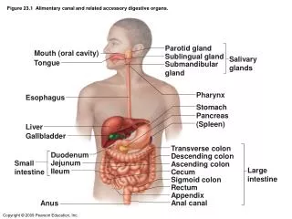
Figure 23.1 Alimentary canal and related accessory digestive organs.
Figure 23.1 Alimentary canal and related accessory digestive organs. Parotid gland. Mouth (oral cavity). Sublingual gland. Salivary glands. Tongue. Submandibular gland. Pharynx. Esophagus. Stomach. Pancreas. (Spleen). Liver. Gallbladder. Transverse colon. Duodenum.
3.28k views • 37 slides

1.7k views • 103 slides
Digestive System: Overview
Digestive System: Overview. The alimentary canal or gastrointestinal (GI) tract digests and absorbs food Alimentary canal – mouth, pharynx, esophagus, stomach, small intestine, and large intestine Accessory digestive organs – teeth, tongue, gallbladder, salivary glands, liver, and pancreas.
1.67k views • 109 slides

Chapter 11, ENDOCRINE SYSTEM
Chapter 11, ENDOCRINE SYSTEM. Section 1 Introduction I. Organization of Endocrine System The functions of the body are regulated by the nervous and the endocrine system. The endocrine system consists of endocrine glands and cells that secrete hormones in various tissues.
1.95k views • 138 slides
Chapter 24 The Digestive System
Chapter 24 The Digestive System. Structures and functions, fig 24.1. Digestive System Organs Alimentary Canal Accessory Digestive Organs Digestive Processes Ingestion Secretion Propulsion Digestion Absorption Defecation. Anatomy of the Digestive System. ________________
1.62k views • 105 slides

IMAGES
COMMENTS
Aug 4, 2021 · 4. Mouth • Opening of the GI tract : receives food, tastes it and prepares it for swallowing • The average volume of the adult mouth is 72 ml in men and …. ml in women • 55 • The mouth is lined by mucous membranes and consists of two major regions: – Vestibule – the space between the inner surface of the cheeks/lips and the teeth – Oral cavity proper – the space inside the ...
Mar 28, 2017 · The digestive system ppt - Download as a PDF or view online for free. ... absorbed in the small intestine, and waste is ejected. The mouth, esophagus, stomach, liver ...
4 The Digestive System Generally, the food moves in one direction and different parts are responsible for doing different jobs in the digestion process. Animation 5 Principle parts of the Alimentary Canal Mouth: physical breakdown of food; tasting; secretion from salivary glands ( saliva ) Saliva: chemical breakdown of carbohydrates
5 Digestive System Anatomy Mouth Journey begins! Teeth: 32 small but hard organs. Made up of dentin and covered in a layer of enamel which is the hardest substance in the body. Living organs that cut and grind food into smaller pieces.
Digestive System * * * A good way to describe peristalsis is an ocean wave moving through the muscle. These diagrams don’t separate the esophagus from the mouth functions, you might want to talk about what happens in the mouth too. * The stomach takes around 4 hours to do it’s job on the food, depending on what kinds of food are digested.
Jul 12, 2024 · The presentation should include: Introduction * Brief overview of the digestive system's role * Importance of understanding its anatomy Major Organs and Structures Oral cavity: Teeth, tongue, salivary glands Pharynx and esophagus: Structure and function in swallowing Stomach: Anatomy, including cardia, fundus, body, antrum, pylorus Small ...
The Processes of Digestion 1. Ingestion taking food into the mouth 2. Secretion GI tract and accessory cells secrete water, acid, buffers, and enzymes into lumen 3. Mixing and Propulsion alternating contraction and relaxation of smooth muscles in the walls of the GI tract 4. Digestion Breaking down of larger food particles into smaller molecules Mechanical Digestion Chemical Digestion 5 ...
The Digestive System CA 5th Grade Science Standards: 5LS2c. students know the sequential steps of digestion and the role of teeth and the mouth, esophagus, stomach, small intestine, large intestine, and colon in the function of the digestive system.
Apr 4, 2023 · The mouth is an important part of the digestive process. Digestion begins in a person’s mouth, which breaks down food into smaller particles. Once a person has finished chewing, their food can ...
Mar 11, 2019 · Digestive System: Overview. Digestive System: Overview. The alimentary canal or gastrointestinal (GI) tract digests and absorbs food Alimentary canal – mouth, pharynx, esophagus, stomach, small intestine, and large intestine Accessory digestive organs – teeth, tongue, gallbladder, salivary glands, liver, and pancreas. 1.67k views • 109 slides